Arrowhead Regional Medical Center (First Floor) Located on the first floor, the Medical Center's state-of-the-art Medical
Imaging Department is completely digital and provides an essential component
of patient care. Medical imaging is considered the "eyes" of
medicine, providing an inside look at a patient's anatomy to help physicians
provide appropriate care.
Address: 400 N. Pepper Ave., Colton, CA 92324
In-Patient Care/Emergency Department Hours: 24 hours, 7 days a week
Additional Locations ARMC also provides medical imaging services at our Arrowhead Family Health
Centers. Please call 855-422-8029 or click the links below to learn more.
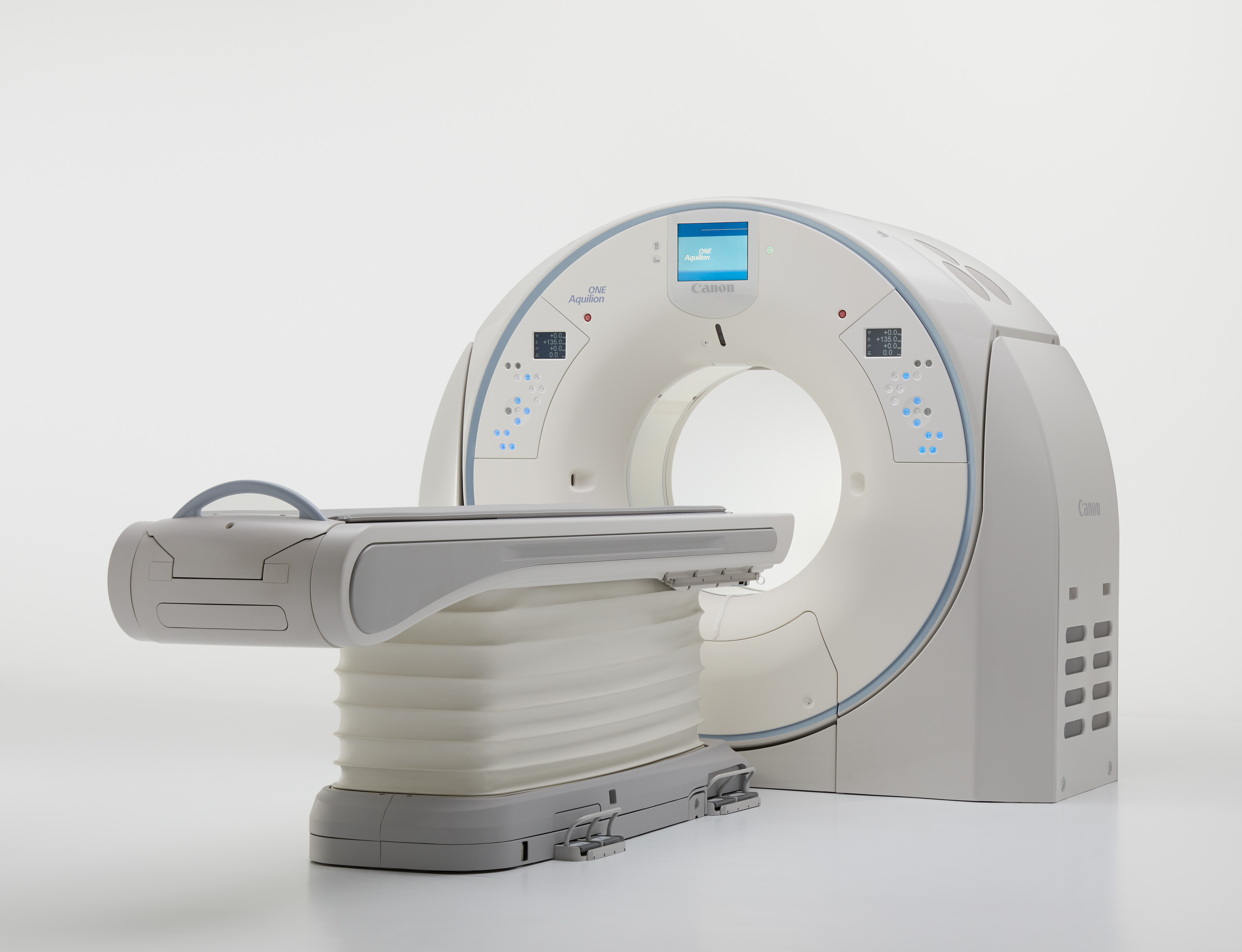
ARMC is proud to introduce
three state-of-the-art Canon CT systems — the Aquilion ONE, Aquilion Prime, and Aquilion Prism — that
enhance imaging quality, speed, and patient comfort. These advanced scanners
deliver high-resolution images with lower radiation doses, faster scan
times, and improved clinical precision to support accurate diagnoses and
better patient outcomes. With innovative technology designed for efficiency
and safety, these new CT systems strengthen our commitment to providing
leading-edge imaging services and exceptional care to our community.
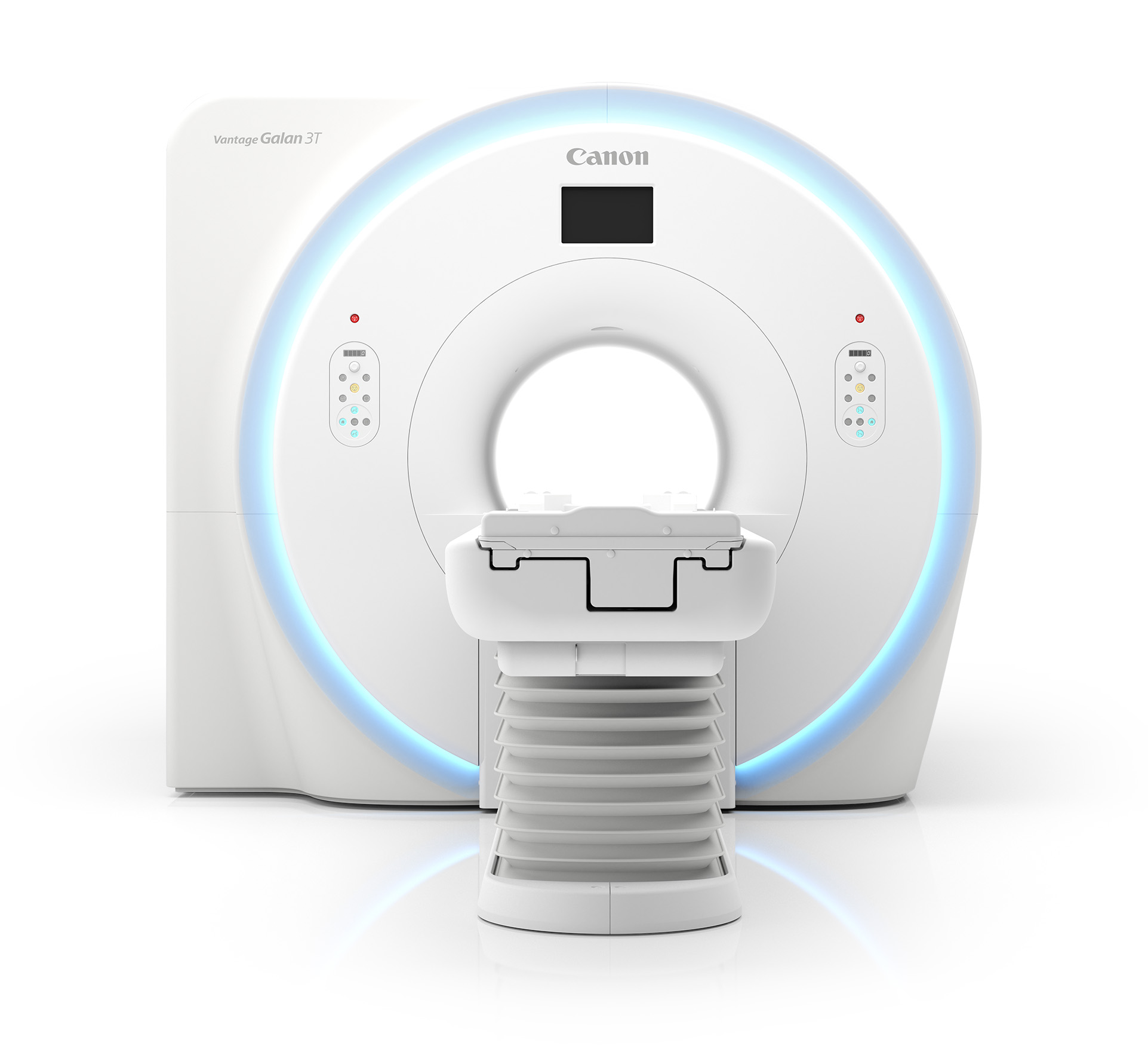
MRI
ARMC is pleased to feature
two advanced Canon MRI systems — the 3T Vantage Galan and the 1.5T Vantage — designed to
elevate diagnostic imaging with exceptional clarity, precision, and patient
comfort. These systems provide high-quality images across a wide range
of clinical applications while reducing scan times and minimizing patient
anxiety through quieter operation and more spacious design. By integrating
these innovative MRI technologies into our imaging services, we reinforce
our commitment to accurate diagnosis, efficient care delivery, and an
improved patient experience.
Other services include:
Radiation oncology with Image Guided Intensity Modulated Radiotherapy (I.G.IMRT),
a precise method of external beam radiation therapy that delivers high
doses of radiation directly to the tumor while sparing the surrounding
healthy tissue.
Two interventional radiology labs for special procedures such as stents,
angiograms, angioplasties and other interventional radiology procedures.
Bone densitometry low-dose system.
Two state-of-the-art digital mammography units and available supine mammography
biopsy unit for patient comfort (mobile service).
Two nuclear medicine units, including one for hearts.
Ultrasound department.
Diagnostic imaging exam services done with state-of-the-art computed radiography
or direct capture system.
State-of-the-art (PACS) Picture Archiving Communication System. Medical
Imaging is completely filmless.
Medical Imaging is staffed by all board certified radiologists (M.D.s)
and all registered and certified technologists in every modality.
American College of Radiology accreditation in Mammography, CT, Nuclear
Medicine, MRI, and Ultrasound.
Speak Up! X-rays, MRIs and Other Medical Imaging Tests
X-rays
What is it? Uses a small amount of radiation to take pictures inside your body.
Used for? Diagnosing broken bones, pneumonia, dental problems. Mammograms are a
common type of X-ray used to help diagnose breast cancer.
What happens? You may be asked to lie still on an X-ray table or sit or stand by the
table. You may wear a lead apron to protect certain parts of your body.
Fact: The amount of radiation you get from an X-ray is small. A chest X-ray
gives out a radiation dose similar to the amount of radiation you are
naturally exposed to from the environment over 10 days.
Ultrasound
What is it? Uses sound waves to create an image. Does not expose you to radiation.
Used for? Diagnosing conditions of the heart, blood vessels, kidneys, liver, and
other organs. During pregnancy, a health care provider uses an ultrasound
to look at the baby.
What happens? You lie on a table. The person giving the test places gel and a device
called a transducer on your skin. The transducer sends out sound waves
that bounce off tissues inside your body.
CT or CAT Scan (Computed Tomography)
What is it? Uses special X-ray equipment to take pictures that show a “slice”
of your body.
What happens? You lie still on a table and may have to hold your breath for a short
time. The CT machine is aimed at the part of your body the health care
provider needs to see. For some CT scans you may receive a “contrast
dye,” which makes parts of your body show up better. The dye may
be given through an intravenous (IV) tube or a syringe in your arm. Some
dye is given in a drink.
MRI (Magnetic Resonance Imaging)
What is it? Uses a large magnet and radio waves to look inside your body. Does not
expose you to radiation.
Used for? Diagnosing torn ligaments, tumors, brain or spinal cord conditions, examining organs.
What happens? You lie still on a table that slides inside a tunnel-shaped machine. You
may have to hold your breath for parts of the exam. For some MRI scans
you may receive a “contrast dye,” which makes parts of your
body show up better. The dye can be given through an intravenous (IV)
tube or a syringe in your arm. Some dye is given in a drink.
Tell your health care provider if you fear small or enclosed spaces, or
if you have:
Metal in your body, such as shrapnel, a bullet, artificial joints or stents
Electronic devices in your body, such as a cardiac pacemaker or implanted pump
Body piercings with metal that cannot be removed
Ever been a welder
Nuclear scans
What is it? Uses radioactive substances and a special camera to see inside your body.
These scans can show how organs, such as your heart and lungs, are working.
What happens? Before the test, you receive a small amount of radioactive material, which
makes parts of your body show up better. The material can be given through
an intravenous (IV) tube or a syringe in your arm. Some is given in a
drink and sometimes you inhale it. You wait as the material is absorbed
by your body. This may take an hour or more. Then you lie still on a table
while the camera takes images.

 ARMC is proud to introduce
three state-of-the-art Canon CT systems — the Aquilion ONE, Aquilion Prime, and Aquilion Prism — that
enhance imaging quality, speed, and patient comfort. These advanced scanners
deliver high-resolution images with lower radiation doses, faster scan
times, and improved clinical precision to support accurate diagnoses and
better patient outcomes. With innovative technology designed for efficiency
and safety, these new CT systems strengthen our commitment to providing
leading-edge imaging services and exceptional care to our community.
ARMC is proud to introduce
three state-of-the-art Canon CT systems — the Aquilion ONE, Aquilion Prime, and Aquilion Prism — that
enhance imaging quality, speed, and patient comfort. These advanced scanners
deliver high-resolution images with lower radiation doses, faster scan
times, and improved clinical precision to support accurate diagnoses and
better patient outcomes. With innovative technology designed for efficiency
and safety, these new CT systems strengthen our commitment to providing
leading-edge imaging services and exceptional care to our community.
 ARMC is pleased to feature
two advanced Canon MRI systems — the 3T Vantage Galan and the 1.5T Vantage — designed to
elevate diagnostic imaging with exceptional clarity, precision, and patient
comfort. These systems provide high-quality images across a wide range
of clinical applications while reducing scan times and minimizing patient
anxiety through quieter operation and more spacious design. By integrating
these innovative MRI technologies into our imaging services, we reinforce
our commitment to accurate diagnosis, efficient care delivery, and an
improved patient experience.
ARMC is pleased to feature
two advanced Canon MRI systems — the 3T Vantage Galan and the 1.5T Vantage — designed to
elevate diagnostic imaging with exceptional clarity, precision, and patient
comfort. These systems provide high-quality images across a wide range
of clinical applications while reducing scan times and minimizing patient
anxiety through quieter operation and more spacious design. By integrating
these innovative MRI technologies into our imaging services, we reinforce
our commitment to accurate diagnosis, efficient care delivery, and an
improved patient experience.
 RadiologyView Profile

